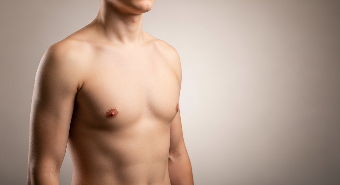

Gynaecomastia, the enlargement of male breast tissue, can cause significant psychological distress and social discomfort during adolescence and adulthood. The MELT (Minimal Excision and Liposuction Technique) represents a surgical advancement designed to address this condition with minimal scarring through a combination of liposuction and targeted tissue excision via a single small incision.
Understanding the MELT Technique
What is the MELT Technique?
The MELT technique is a surgical procedure specifically developed to treat gynaecomastia, combining subcutaneous liposuction with direct glandular excision through a single periareolar incision. This minimally invasive approach was created to reduce scarring while effectively removing both excess fatty tissue and fibrous glandular tissue.
How Does MELT Differ from Traditional Approaches?
Traditional gynaecomastia surgery typically requires separate or larger incisions for liposuction and tissue removal. The MELT technique integrates both procedures through one small incision at the border of the areola, where scarring is less visible. This consolidated approach reduces surgical trauma, operative time, and the number of tissue entry points.
The MELT Procedure: Step-by-Step
Pre-Operative Assessment
Before the procedure, the surgeon evaluates the extent of gynaecomastia through physical examination and may order imaging studies. The assessment determines the ratio of fatty to glandular tissue, which influences the surgical approach. Patients undergo standard pre-surgical testing, including blood work and medical clearance.
Surgical Technique
Anesthesia Administration
The procedure is typically performed under general anaesthesia or local anaesthesia with sedation, depending on the extent of tissue involved.
Incision Placement
A single incision is made along the inferior border of the areola, typically measuring 2-3 centimetres. This location provides access to both liposuction and tissue excision while concealing the scar within the natural colour transition.
Liposuction Phase
Through the periareolar incision, a cannula is inserted to perform subcutaneous liposuction. The surgeon removes excess fatty tissue using either traditional suction-assisted or power-assisted liposuction techniques. This step reduces the overall breast volume and facilitates access to the glandular tissue.
Glandular Excision
Following liposuction, the surgeon directly excises the firm glandular tissue through the same incision. Careful attention is paid to preserving the nipple-areolar complex and maintaining adequate tissue to maintain proper chest contour. A small amount of glandular tissue is typically left beneath the areola to prevent nipple retraction.
Closure
The incision is closed in layers using absorbable sutures. A compression garment is applied immediately to minimise swelling and support tissue adherence to the chest wall.
Duration and Recovery Timeline
The MELT procedure typically takes 1-2 hours, depending on the extent of tissue involvement. Most patients are discharged the same day with specific post-operative instructions.
Expected Outcomes and Results
Cosmetic Results
The MELT technique produces a flatter, more masculine chest contour with minimal visible scarring. Because the incision is placed at the areolar border, it becomes nearly invisible as healing progresses. Most patients see significant improvement in chest appearance within 3-6 months as swelling resolves and tissues settle.
Functional Benefits
Beyond appearance, the MELT technique offers several functional advantages:
- Reduced surgical trauma: Single-incision approach minimises tissue damage
- Faster recovery: Combined procedure reduces overall operative time and healing period
- Lower infection risk: Fewer incisions mean fewer potential entry points for bacteria
- Improved tissue adherence: Simultaneous liposuction and excision allow for better skin retraction
Scarring and Aesthetic Considerations
Scarring from the MELT technique is typically limited to a thin line at the inferior areolar border. The scar fades over 12-18 months and is camouflaged by the natural pigment change between the areola and surrounding skin. Proper wound care and scar management can further minimise visibility.
Potential Complications and Risks
Common Post-Operative Effects
Swelling and Bruising
Temporary swelling and bruising are normal and typically resolve within 2-4 weeks. Compression garments help minimise these effects.
Numbness
Temporary numbness of the nipple or chest skin may occur due to nerve trauma but usually resolves within several months.
Asymmetry
Minor asymmetry may be present as swelling resolves unevenly, though this typically improves with complete healing.
Less Common Complications
Hematoma or Seroma
Blood or fluid collections may develop postoperatively and require drainage in some cases.
Contour Irregularities
Over-resection or under-resection of tissue can create surface irregularities. Revision surgery may be necessary if these do not resolve.
Nipple-Areolar Complex Changes
Though rare, changes in nipple sensation, position, or appearance can occur.
Infection
Surgical site infections occur in less than 2% of cases when proper sterile technique and post-operative care are followed.
Recovery and Post-Operative Care
Immediate Post-Operative Period (Days 1-7)
Patients wear compression garments continuously during the first week. Pain is typically mild to moderate and controlled with prescribed medications. Most individuals return to light activities within 3-5 days, though strenuous activity remains restricted.
Subacute Recovery (Weeks 2-6)
Compression garments are continued for 4-6 weeks to support healing and reduce swelling. Patients gradually resume normal activities, avoiding heavy lifting and intense exercise until cleared by the surgeon, typically between weeks 4 and 6.
Long-Term Healing (Months 3-12)
Swelling typically decreases over several months, with final results evident around 6-12 months post-surgery. Scar maturation continues for up to 18 months, during which scars fade and become less noticeable.
Activity Restrictions
- Week 1: Light walking only, no lifting over 5 pounds
- Weeks 2-4: Gradual return to normal daily activities, continued avoidance of strenuous exercise
- Weeks 4-6: Progressive reintroduction of upper body exercises as cleared by the surgeon
- After 6 weeks: Return to full activity, including weightlifting and contact sports
Who is an Ideal Candidate?
Patient Selection Criteria
The MELT technique works for patients with:
- Grade I or II gynaecomastia: Mild to moderate breast enlargement without significant skin excess
- Mixed tissue composition: A combination of fatty and glandular tissue
- Good skin elasticity: Adequate skin quality for retraction after tissue removal
- Stable weight: Patients at or near their ideal body weight
- Realistic expectations: Understanding of achievable outcomes and potential limitations
Contraindications
The MELT technique may not be suitable for:
- Severe gynaecomastia with excess skin: May require additional skin excision procedures
- Predominantly fibrous tissue: Dense glandular tissue may be difficult to remove through a limited incision
- Active infections or uncontrolled medical conditions: Must be resolved before surgery
- Unrealistic expectations: Patients expecting perfection or immediate results
Alternative Treatment Options
Liposuction Alone
For patients with primarily fatty gynaecomastia and minimal glandular tissue, liposuction without excision may be sufficient. This approach uses even smaller incisions but cannot address firm glandular tissue.
Traditional Excision
Patients with predominantly glandular tissue or severe gynaecomastia may require traditional surgical excision, which can involve larger incisions or additional procedures to remove excess skin.
Ultrasound-Assisted or Laser-Assisted Techniques
Some surgeons combine the MELT approach with ultrasound-assisted liposuction (UAL) or laser-assisted techniques to improve fat removal and potentially enhance skin tightening.
Non-Surgical Options
Mild cases of gynaecomastia related to weight gain may respond to weight loss and exercise. However, true glandular tissue typically requires surgical intervention for removal.
Cost Considerations
The cost of the MELT technique varies based on geographic location, surgeon experience, and facility fees. In the United States, the procedure typically costs between $4,000 and $8,000. This cost usually includes:
- Surgeon’s fees
- Anesthesia fees
- Facility or operating room fees
- Post-operative garments
- Follow-up appointments
Insurance coverage for gynaecomastia surgery is limited and typically requires documentation that the condition causes physical symptoms (such as pain) rather than purely cosmetic concerns.
Conclusion
The MELT technique offers an effective surgical solution for gynaecomastia, with reduced scarring and a shorter recovery time. The single-incision approach addresses both fatty and glandular components while minimising visible marks.
If you are experiencing breast enlargement, tenderness, or psychological distress related to chest appearance, consult with a board-certified plastic surgeon experienced in gynaecomastia treatment to determine if the MELT technique is appropriate for your condition.
