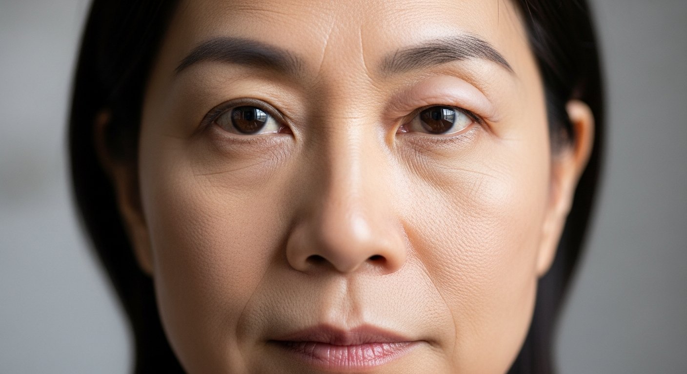

Can you close your eyes completely without effort? Droopy eyelids fall into two distinct medical categories that require different treatment approaches. Ptosis, which involves the actual eyelid muscle and its ability to lift the lid, and dermatochalasis, which refers to excess skin that accumulates on the upper eyelid over time.
Both conditions can affect vision and facial appearance, but their underlying mechanisms differ substantially. Understanding which condition you have—or whether you have both—determines the appropriate surgical approach and expected outcomes.
Ptosis
Ptosis occurs when the levator muscle—the primary muscle responsible for lifting the upper eyelid—loses its ability to elevate the lid adequately. The levator muscle attaches to the tarsal plate of the eyelid through an aponeurosis (a thin, fibrous tissue that connects muscle to bone). When this attachment stretches, detaches, or when the muscle itself weakens, the eyelid droops.
Measuring Ptosis Severity
Doctors who specialise in eye conditions and plastic surgeons measure ptosis using several clinical parameters:
- Marginal reflex distance (MRD1): The distance from the upper eyelid margin to the corneal light reflex (the reflection of light on the front surface of the eye).
- Levator function: How far the eyelid travels from downgaze to upgaze (this measures how well the lifting muscle is working).
- Palpebral fissure height: The vertical opening between the upper and lower lids.
Types of Ptosis
Aponeurotic ptosis represents a commonly seen form in adults. The levator aponeurosis (the connective tissue linking the muscle to the eyelid) stretches or pulls away from its attachment. This often results from age-related tissue changes, contact lens wear, or previous eye surgery. The levator muscle itself functions normally, but the force transmission to the eyelid becomes inefficient.
Myogenic ptosis involves the levator muscle directly. Conditions such as myasthenia gravis (a condition in which muscles become weak), chronic progressive external ophthalmoplegia (a condition causing gradual weakening of eye muscles), or muscular dystrophy affect muscle function. This type often shows variable drooping throughout the day or with fatigue.
Neurogenic ptosis results from nerve damage affecting the oculomotor nerve (the third cranial nerve, which controls eye movement) or the sympathetic pathway (Horner syndrome—a condition affecting the nerve pathway from the brain to the face and eye). Third-nerve palsy causes severe ptosis and associated eye movement abnormalities. Horner syndrome produces milder ptosis, pupil constriction, and reduced facial sweating.
Mechanical ptosis occurs when eyelid masses, scarring, or inflammation weigh down or restrict the lid.
Dermatochalasis
Dermatochalasis develops when the thin eyelid skin loses elasticity and forms folds. Unlike ptosis, the levator muscle (the eyelid-lifting muscle) functions normally. The eyelid itself opens fully, but redundant skin hangs over the lid margin.
Contributing Factors
The eyelid contains some of the thinnest skin on the body. This delicate tissue responds to:
- Collagen degradation: Ultraviolet exposure breaks down collagen and elastin fibres (the proteins that give skin its structure and elasticity) over decades
- Gravitational effects: Skin gradually stretches and descends with age
- Orbital fat prolapse: Fat pads behind the orbital septum (the membrane separating the eyelid from deeper structures) push forward, creating fullness
- Genetic predisposition: Some individuals develop excess skin earlier than others
- Lifestyle factors: Smoking accelerates skin ageing. Chronic eye rubbing stretches tissue.
Clinical Presentation
Patients with dermatochalasis describe a “heavy” sensation in their upper eyelids, particularly towards the end of the day. The excess skin may touch the eyelashes or obstruct the superior visual field. You can manually lift the redundant skin away from the eye. This immediately improves the visual field. This manoeuvre helps distinguish dermatochalasis from true ptosis.
Distinguishing Between the Two Conditions
Clinical examination differentiates ptosis from dermatochalasis through several assessments:
Eyelid crease position provides valuable information. In ptosis, the crease often sits higher than normal or appears absent because the levator aponeurosis (the connective tissue of the lifting muscle) has detached from its usual position. In dermatochalasis, the crease remains in its normal position but becomes hidden beneath skin folds.
Upper lid margin position relative to the pupil differs between conditions. Ptosis lowers the lid margin, potentially obscuring the pupil. Dermatochalasis keeps the margin in the normal position despite the overhanging skin.
Frontalis compensation occurs when patients unconsciously raise their eyebrows to lift droopy lids. Both conditions can trigger this compensation, but the degree often correlates with functional severity.
💡 Did You Know?
The levator muscle performs numerous blinks daily, making it one of the most active muscles in the body. This constant use explains why aponeurotic stretching (the gradual pulling away of the muscle’s connective tissue) develops gradually over decades.
Combined Presentations
Many patients seeking evaluation for droopy eyelids have both ptosis and dermatochalasis simultaneously. Age-related changes affect multiple structures concurrently—the levator aponeurosis (muscle connective tissue) stretches while the skin loses elasticity. Addressing only one component may leave the others untreated, affecting the overall outcome.
Surgical planning for combined cases requires treating both elements. Removing excess skin alone when ptosis is present leaves the patient with persistent lid drooping despite the skin removal. Conversely, repairing the muscle problem without addressing significant excess skin may still leave redundant skin affecting appearance and function.
Surgical Approaches for Ptosis
Ptosis surgery addresses the underlying muscular or connective tissue problem rather than simply removing skin. The surgical technique depends on levator function measurements (how well the eyelid-lifting muscle works):
External Levator Advancement
For patients with good levator function, the surgeon makes an incision along the eyelid crease to directly see and reattach the stretched connective tissue. The surgeon identifies the levator, advances it to the tarsal plate (the firm tissue within the eyelid), and secures it at the appropriate tension. The surgeon makes adjustments during surgery to fine-tune lid height and contour.
Müller Muscle-Conjunctival Resection
This internal approach works for mild ptosis with good levator function. Operating from behind the eyelid, the surgeon removes a calculated amount of Müller muscle (a secondary eyelid-lifting muscle) and conjunctiva (the clear membrane covering the white of the eye). The surgeon performs phenylephrine testing beforehand to predict the response to this procedure. Patients whose lids elevate with these drops typically respond well.
Frontalis Suspension
Severe ptosis with poor levator function requires attaching the eyelid to the frontalis (forehead) muscle using a sling material. When the patient raises their eyebrows, the eyelid elevates. Materials include silicone rods, expanded polytetrafluoroethylene, or autogenous fascia lata (tissue harvested from the patient’s own thigh).
Surgical Approaches for Dermatochalasis
Upper blepharoplasty (eyelid surgery to remove excess skin) removes excess skin and, when present, prolapsed orbital fat (fat that has pushed forward). The procedure involves the following steps:
- Marking the excision: The surgeon marks the lower incision line at the natural eyelid crease. The upper line determines how much skin to remove. Pinch testing ensures adequate closure without lagophthalmos (inability to fully close the eye).
- Skin excision: The surgeon removes a thin strip of skin. A thin strip of orbicularis oculi (the muscle that closes the eyelid) may be included during skin removal to prevent a thick, rounded appearance.
- Fat management: The surgeon conservatively reduces or repositions prolapsed medial and central fat pads. Over-removal creates a hollow, aged appearance.
- Closure: The surgeon places fine sutures carefully to minimise visible scarring.
⚠️ Important Note
Conservative skin removal prevents lagophthalmos, a condition in which the eyelid cannot fully close. This complication causes corneal exposure (when the front surface of the eye is exposed to air), dryness, and potential vision-threatening complications.
Functional vs Aesthetic Considerations
Both conditions may qualify for medical necessity when they impair vision. Visual field testing (a diagnostic test that measures how much you can see in your peripheral vision) documents the degree of superior field obstruction. Your healthcare provider can discuss whether this technology might be suitable for your situation based on your individual symptoms and test results.
Functional indicators include:
- Superior visual field loss on formal testing
- Difficulty reading or performing tasks requiring upward gaze
- Compensatory head posturing (tilting your chin up to see beneath droopy lids)
- Eye fatigue from chronic frontalis muscle activation (constantly raising your eyebrows to lift the lids)
Aesthetic motivations are considered alongside functional indications during the pre-operative assessment process.
Recovery Expectations
Ptosis surgery and blepharoplasty share similar recovery timelines:
First week: Swelling and bruising peak at days 2-3, then gradually resolve. Cold compresses and head elevation help. The surgeon typically removes sutures (stitches) on days 5-7.
Weeks 2-4: Residual swelling diminishes. The eyelids may appear asymmetric during this period as swelling resolves unevenly.
Months 1-3: Final contour and lid position stabilise. Minor asymmetries often self-correct as tissues settle.
You should initially avoid activities including strenuous exercise, bending forward, and anything that increases blood flow to the face.
Putting This Into Practice
- Obtain a comprehensive eye examination, including visual acuity (a test measuring how clearly you see), visual field testing (a diagnostic test measuring peripheral vision), and assessment for dry eye or other ocular surface conditions that might affect healing.
- Discontinue blood-thinning substances for the recommended period before surgery. This includes prescription anticoagulants (medications that prevent blood clotting—only discontinue with your prescribing physician’s guidance), aspirin, non-steroidal anti-inflammatory drugs (such as ibuprofen), vitamin E, fish oil, and certain herbal supplements.
- Arrange post-operative support for the first 24-48 hours. Vision may be blurred from the ointment. Driving is not advisable immediately after surgery.
- Prepare your recovery space with prescribed eye drops, clean gauze, cold compresses, and entertainment that doesn’t require extensive reading.
- Document the current appearance with photographs in good lighting, with the subject looking straight ahead and with relaxed facial muscles.
When to Seek Professional Help
- Sudden onset of eyelid drooping, particularly with headache, pupil changes, or double vision
- Progressive worsening of lid position over weeks rather than years
- Drooping that varies throughout the day or worsens with fatigue
- Eyelid drooping following trauma or eye surgery
- Associated symptoms such as difficulty swallowing, limb weakness, or breathing changes
- Significant visual field obstruction affecting daily activities
- Asymmetry developing in previously symmetric eyelids
Commonly Asked Questions
How do I know if I need ptosis surgery or blepharoplasty?
Clinical examination determines which condition predominates. If manually lifting the excess skin fully corrects the drooping, dermatochalasis (excess skin) is the primary issue. If the lid margin remains low despite skin elevation, ptosis (muscle weakness) exists. Many patients require both procedures performed together for complete correction. Your doctor will assess your specific situation to determine the most appropriate course of action.
Can my eyelids look natural after ptosis surgery?
Surgeons aim for natural-appearing results with appropriate lid height, contour, and symmetry. The goal is restoration of normal anatomy rather than creation of an artificial appearance.
Can ptosis recur after surgery?
Aponeurotic ptosis repair generally aims for durable correction, though gradual tissue changes may produce some recurrence over many years.
Patients with myogenic (muscle-based) or neurogenic (nerve-based) causes may experience progression related to their underlying condition rather than surgical failure.
Is the surgery performed under local or general anaesthesia?
Most ptosis and blepharoplasty procedures use local anaesthesia (numbing medication injected into the area) with sedation (medication to help you relax). Patients remain comfortable while the surgeon can assess lid position with the patient sitting upright. Pure local anaesthesia is possible for patients who prefer minimal sedation.
What are the risks specific to ptosis surgery?
Under-correction (the lid remains too low) or over-correction (the lid sits too high) may require revision surgery. Asymmetry, though usually minor, can occur. Lagophthalosis (incomplete lid closure) typically resolves as swelling settles, but occasionally persists. Dry eye symptoms (such as grittiness or burning) may temporarily worsen before improving.
Conclusion
Clinical examination determines whether ptosis, dermatochalasis, or both are present in your eyelids. Accurate diagnosis guides surgical planning—ptosis requires repair of the levator muscle or connective tissue, while dermatochalasis requires skin removal. Visual field testing documents functional impairment when present.
If you’re experiencing droopy eyelids, incomplete eye closure, or visual field obstruction, a plastic surgeon can help determine which condition is present and whether any intervention is appropriate.
