

Ptosis occurs when the upper eyelid sits lower than its normal position. It may result from abnormal development of the eyelid-lifting muscle, stretching or detachment of its connective-tissue attachment, nerve dysfunction, muscle disease or mechanical factors affecting the eyelid. The underlying tissue abnormality commonly differs between these two forms, and that difference typically helps determine which surgical technique is appropriate and what outcomes are realistic.
In simple congenital ptosis, the levator muscle is commonly underdeveloped and may contain increased fibrous or fatty tissue, resulting in reduced contraction and relaxation. In the most common acquired form, the levator muscle itself remains relatively functional, but its aponeurotic attachment has stretched, thinned or detached. Other acquired forms may result from nerve, muscle, trauma or mechanical causes. In acquired ptosis, the muscle structure was once normal, meaning the same degree of droop may respond to a very different repair.
Understanding which type is present generally requires measuring specific clinical parameters, including levator function, margin reflex distance, lid crease position, and fatigue response, before any surgical planning can begin.
Congenital Ptosis: Characteristics and Causes
Congenital ptosis appears at birth or within the first year of life. The primary cause is often dysgenesis of the levator muscle: the muscle fibres can fail to develop properly and are frequently replaced by fibrous fatty tissue. This abnormal tissue may not contract effectively, potentially resulting in reduced eyelid elevation.
Children with congenital ptosis typically show a characteristic pattern: the affected eyelid sits lower than normal when looking straight ahead. When the child looks down, the affected eyelid may remain relatively higher than the other eyelid. This is known as lid lag on downgaze and reflects the reduced ability of the abnormal levator muscle to relax and lengthen normally. This “lid lag” occurs because the fibrotic muscle tissue frequently cannot relax fully either.
Types of Congenital Ptosis
Simple congenital ptosis involves only the levator muscle. The condition may be unilateral or bilateral, with severity ranging from mild cosmetic concerns to complete obstruction of the visual axis.
Blepharophimosis syndrome combines ptosis with other eyelid abnormalities, including telecanthus (increased distance between inner eye corners), epicanthus inversus (skin folds below the inner corners), and shortened horizontal eyelid length. This genetic condition typically requires staged surgical correction addressing multiple structural issues.
Marcus Gunn jaw-winking ptosis involves aberrant nerve connections between the muscles controlling jaw movement and eyelid elevation. The ptotic eyelid may elevate during chewing or lateral jaw movement, a distinctive finding that helps establish the diagnosis and influences the surgical approach.
Visual Development Concerns
In children, moderate to severe congenital ptosis poses risks beyond cosmetic appearance. Congenital ptosis can affect visual development by obstructing the visual axis or by contributing to astigmatism, unequal focusing between the eyes or strabismus. These problems may lead to amblyopia if they are not detected and treated early.
Children may adopt a persistent chin-up head posture to see beneath the eyelid. This can cause postural strain and may interfere with daily activities and social interaction. Early surgical intervention may be recommended when ptosis threatens visual development, even before the ideal surgical age.
Acquired Ptosis: Types and Mechanisms
Acquired ptosis develops after normal eyelid function has been established. Identifying the underlying mechanism guides both workup and treatment decisions.
Aponeurotic Ptosis
A frequent form of acquired ptosis involves stretching or dehiscence of the levator aponeurosis, the thin tissue connecting the levator muscle to the eyelid. Age-related tissue changes, chronic eye rubbing, contact lens wear over many years, or previous eye surgery can weaken this connection.
Aponeurotic ptosis commonly presents with a high or poorly defined eyelid crease, relatively good levator function (the muscle works but isn’t properly connected), and normal eyelid movement when looking down. These features distinguish it from other acquired forms and may support a favourable surgical prognosis with aponeurosis repair techniques.
Neurogenic Ptosis
Damage to the nerves controlling eyelid elevation can cause neurogenic ptosis. Third nerve (oculomotor) palsy typically produces ptosis accompanied by eye movement abnormalities and pupil changes, findings that require prompt neurological evaluation to rule out an aneurysm or other serious causes.
Horner syndrome involves disruption of sympathetic nerve pathways, causing mild ptosis combined with pupil constriction and sometimes reduced facial sweating. The ptosis is typically subtle because only the Müller muscle (a secondary eyelid elevator) is affected.
Myogenic Ptosis
Conditions affecting muscle tissue itself can cause myogenic ptosis. Myasthenia gravis produces variable ptosis that worsens with fatigue and improves with rest; the ptosis may be minimal in the morning and severe by evening. Chronic progressive external ophthalmoplegia and other mitochondrial disorders cause slowly progressive ptosis, often accompanied by limited eye movements.
Mechanical Ptosis
Eyelid tumours, inflammation, swelling or scarring can physically weigh down or restrict the eyelid. Excess upper-eyelid skin may create the appearance of ptosis without lowering the eyelid margin, known as pseudoptosis, although both conditions may coexist. Treatment addresses the underlying cause rather than the levator mechanism.
Diagnostic Evaluation
Accurate diagnosis requires a systematic clinical assessment measuring multiple parameters.
Margin reflex distance (MRD1) measures the distance from the upper eyelid margin to the corneal light reflex when looking straight ahead. Normal values range from 4- 5 mm; lower values indicate more significant ptosis.
Levator function assesses how much the eyelid elevates from full downgaze to full upgaze while blocking brow movement. Values above 12mm indicate excellent function, roughly 9 to 11mm indicate good function (typical in aponeurotic ptosis), 5 to 9mm indicate fair function, and 4mm or less indicate poor function (common in congenital ptosis with muscle dysgenesis).
Eyelid crease position and presence help differentiate types. Aponeurotic ptosis often shows a high or absent crease, while congenital ptosis may have an absent or poorly defined crease.
Fatigue testing involves sustained upgaze for several minutes to detect worsening suggestive of myasthenia gravis. Additional tests, including ice pack application and blood work, may follow if myasthenia is suspected.
Did You Know?
Phenylephrine eye drops can temporarily elevate a ptotic eyelid by stimulating the Müller muscle. This test helps predict whether Müller muscle surgery alone might achieve adequate correction or whether levator surgery is necessary.
Surgical Treatment Approaches
Ptosis correction surgery selection depends on ptosis type, severity, and levator function. Each technique is designed to address specific anatomical problems.
External Levator Advancement
The standard approach for aponeurotic ptosis involves an incision in the natural eyelid crease. The surgeon identifies the stretched or dehisced aponeurosis, advances it to a more appropriate position, and secures it to the tarsal plate. This aims to restore the normal mechanical connection between the muscle and the eyelid. This procedure is sometimes combined with upper eyelid surgery when excess eyelid skin is also present.
Adjustments during surgery allow precise height setting. Patients are often asked to open their eyes during the procedure (under local anaesthesia) to verify symmetric eyelid position before final suture placement.
Müller Muscle-Conjunctival Resection
For mild ptosis with good levator function and a positive response to phenylephrine testing, this internal approach removes a measured amount of Müller muscle and overlying conjunctiva through the inner eyelid surface. Visible external scarring is typically avoided.
Frontalis Sling
When the levator function is poor (below 4mm), direct muscle surgery cannot achieve adequate elevation. Frontalis sling procedures connect the eyelid to the brow muscle using either autogenous fascia lata (tissue harvested from the thigh) or synthetic materials.
This technique is frequently used for congenital ptosis with severe muscle dysgenesis. The brow muscle assumes eyelid elevation function, and patients learn to raise their eyebrows to open their eyes. While functional, this creates a different movement pattern than normal eyelid opening.
Recovery and Expectations
Most visible swelling and bruising improve over approximately two to three weeks, although mild residual swelling and changes in eyelid contour may take longer to settle. Initial eyelid position may appear higher than the final result, as swelling settles and tissues relax into their new position. Final assessment of surgical outcome usually occurs around three to six months.
Important Note
Temporary incomplete eyelid closure can occur after ptosis surgery, particularly following larger corrections or procedures performed for poor levator function. Lubrication may be prescribed to protect the ocular surface while healing occurs. Persistent dryness, pain, blurred vision or difficulty closing the eye should be reported promptly.
Residual asymmetry, undercorrection, overcorrection or recurrence may lead to consideration of revision surgery. Perfect symmetry cannot always be guaranteed because the two eyelids may differ in muscle function, anatomy and healing response.
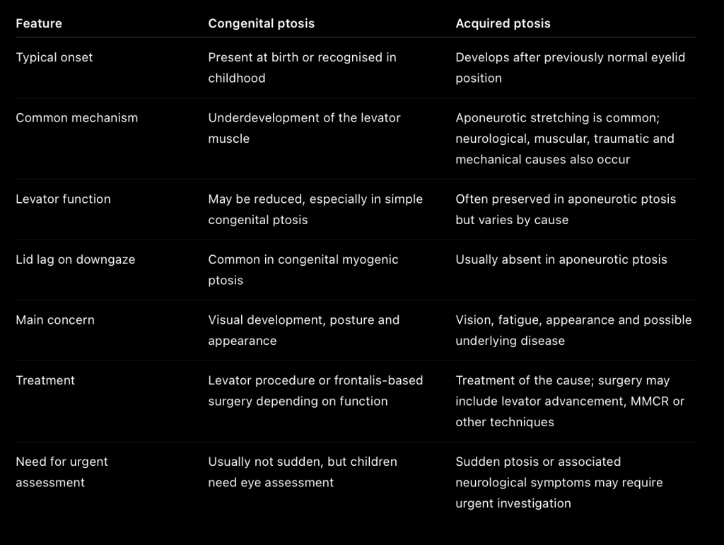
Congenital vs Acquired: Key Differences Summary
When to Seek Professional Help
- Eyelid drooping that develops suddenly, particularly with headache, pupil changes, or double vision
- Progressive worsening of existing ptosis over weeks to months
- Ptosis that varies significantly throughout the day
- Eyelid drooping accompanied by eye redness, pain, or swelling
- Childhood ptosis covering the pupil or causing head tilt
- Ptosis interfering with reading, driving, or daily activities
Commonly Asked Questions
Can ptosis correct itself without surgery?
Simple congenital ptosis usually does not resolve spontaneously because the underlying levator abnormality is structural. Some forms of acquired ptosis may resolve if the underlying cause is treated. For example, ptosis from myasthenia gravis may improve with medical management, and postoperative swelling that causes temporary ptosis typically resolves as the wound heals.
At what age should congenital ptosis be repaired?
Timing depends on severity and is individualised. Surgery may be considered early when the eyelid obstructs vision, induces significant astigmatism or causes a persistent chin-up posture. When vision is not threatened, correction may be delayed until the child is older and more cooperative with examination and postoperative care.
Will ptosis surgery affect my ability to close my eyes?
Some temporary difficulty with complete eye closure is expected after surgery and commonly improves as healing progresses. Surgeons aim to balance adequate eyelid elevation against maintaining the ability to close the eye completely, as incomplete closure risks corneal exposure and dryness.
Is ptosis surgery performed under general or local anaesthesia?
Many adults can undergo ptosis surgery under local anaesthesia, with or without light sedation. General anaesthesia is commonly used for children and may be selected for adults depending on the procedure, medical condition and patient preference.
How long do ptosis surgery results last?
Surgical correction typically provides long-lasting results. However, aponeurotic ptosis can recur years later as tissues continue to age. Frontalis sling procedures may require adjustment or revision as synthetic materials stretch or shift over time.
Next Steps
Treatment selection depends on the underlying cause, severity of ptosis, levator function, eyelid anatomy, ocular-surface health and the patient’s functional and aesthetic goals. Congenital ptosis may be treated with a levator procedure or a frontalis-based technique depending on muscle function. Acquired aponeurotic ptosis commonly responds to repair or advancement of the levator aponeurosis, while neurological and muscular causes may require medical investigation and treatment before eyelid surgery is considered.
Sudden-onset ptosis accompanied by pupil changes or double vision requires prompt neurological evaluation before any surgical planning.
If you are experiencing a drooping upper eyelid, whether present since childhood, worsening gradually with age, or varying throughout the day, consider scheduling a clinical evaluation with an accredited plastic surgeon to review levator function and margin reflex distance to identify an appropriate surgical approach.
